Grocott-Gomori’s Methenamine Silver Staining – Principle, Procedure, Applications
 Biology Notes Online" width="" height="" />
Grocott-Gomori’s Methenamine Silver Staining is a type of special staining technique used in microbiology and histology to visualize fungal organisms, specifically those that form characteristic spores known as arthroconidia.
The staining technique was developed in the 1930s by Grocott and Gomori and is based on the use of silver and methenamine. The fungal cells are stained with a silver nitrate solution, which is then developed by the addition of methenamine to produce a characteristic black-stained pattern that makes the fungal cells easily visible under a microscope.
The importance of Grocott-Gomori’s Methenamine Silver Staining lies in its ability to specifically visualize fungal organisms, which can be difficult to see using other staining techniques. It is especially useful in the diagnosis of fungal infections, where the identification of the causative organism is critical for effective treatment.
The history of Grocott-Gomori’s Methenamine Silver Staining is rooted in the development of microbiology and histology as scientific disciplines, and it represents one of the many innovations in staining techniques that have helped to advance our understanding of the microorganisms that surround us. Today, the staining technique continues to be widely used in clinical and research settings and is an important tool for the diagnosis and study of fungal infections.
What is Grocott-Gomori’s Methenamine Silver Staining?
- The histology stain known as Grocott-methenamine Gomori’s silver (GMS) is most often used to identify carbohydrates in fungal microorganisms.
- Named after the Hungarian physician who pioneered the technique, György Gömöri staining is now widely used.
- It was first used to detect Pneumocystis jiroveci, a fungus that causes pneumocytosis and is found in immunocompromised and immunosuppressed individuals, but its original purpose was to evaluate missing tissues and disorders in the liver and the rectum (Nadworny, Wang, Tredget, & Robert, 2010).
- Goromi is more sensitive to detecting fungi and other polysaccharide-rich microorganisms in paraffin prepared sections than other stains like Periodic Acid-Schiffs stain and Gridley Stain.
- The main reagents in traditional Grocott stain are Gomori’s methenamine-silver nitrate and chromic acid.
- Tissue slices have also been examined using this method to positively identify fungus.
- With its use in Histology, fungus in aspirates, tissues, and smears can be easily detected and displayed.
Objectives
- To prove that fungi are present in a sample.
- Pneumocystis jirovecii and Histoplasma spp. must be shown to exist.
Grocott-Gomori’s Methenamine Silver Staining Principle
Chromic acid oxidation generates aldehydes from polysaccharide components of fungal cell wall, which are proven by the reduction of an alkaline hexamine-silver complex. This reaction is analogous to the periodic acid Schiff reaction (see PAS).
Grocott’s alkaline hexamine-silver solution is a carrier that, upon reduction, precipitates nascent silver ions and thus darkens the site. The term for this is “argentaffin response.”
Argentaffin reaction – the ability of a silver complex solution to blacken tissue without a reducing bath. The term is adjectival and applies to numerous techniques (eg von Kossa). Therefore, the term “argentaffin response” should not be used as a proper noun.
In most developed nations, fungal infections manifesting as opportunistic infections in immunocompromised people are on the rise. Fungi stain poorly with haematoxylin and are PAS-positive due to the presence of carbohydrates in their cell walls.
Important Notes
- A recognised positive control section must be utilised to assure proper distinction.
- The preparation of reagents requires a fume hood.
- Measure the volume of chromic acid using the microwave technique.
- When microwaving solutions, ensure that the lids are not tightly fastened to allow for expansion.
- Before placing slides in microwave-heated staining solutions, ensure that the solutions are completely mixed to provide a uniform temperature distribution.
- Note that removed solutions from the microwave may spontaneously boil.
- Timing may vary based on microwave type and power.
- A combination of the original method and the microwave method may be utilised; for example, the microwave approach for silver may be combined with the original method for chromic acid, or vice versa.
Reagents Requireds
Solution of chromium trioxide, solution of sodium bisulfite, solution of silver nitrate, solution of methenamine, solution of borax, solution of gold chloride, solution of sodium thiosulfate, solution of light green stock.
Solution Preparation
Manual preparation kits are the standard delivery method for these reagents, while some solutions are available ready-to-use. The staining concentrations you’ll need will determine how you prepare the solutions.
- Chromic Acid(1.4%): In order to make chromic acid (1.4%), you need 4 grammes of chromium trioxide and 100 millilitres of distilled water.
- Substitute for silver: silver nitrate 5% in 23ml bottle with 3% methanamine/hexamine 1.25 ml with 5% Borax (Sodium tetraborate) Distiller’s water (3ml) and tap water (25ml)
- 3.2% Sodium Chloroaurate (Yellow gold chloride): Gold chloride (Sodium Chloroaurate, or “Yellow Gold Chloride”), 3.2 percent 1.0g + 500mL of Pure H2O
- 4.2 % Sulfate of sodium (Hypo): 2.0 grammes of sodium sulphate plus 100 millilitres of distilled water
- Working Light Green Stock Solution: Solving for the Problem with Green Light Stock 10 millilitres of light green in 40 millilitres of acetic acid.
- Silver solution: 3% methanamine (hexamine) 23 ml, 5% silver nitrate 1.25 ml, 5% borax (sodium tetraborate) 3 ml, Distilled water 25 ml.
Procedure of Grocott-Gomori’s Methenamine Silver Staining
General Method
- First, immerse the pieces in distilled water.
- Secondly, oxidise with 4% aqueous chromic acid for 1 hour at room temperature.
- Third, rinse for a little period of time.
- Soak the tissue slices in sodium metabisulphite 1% for a full minute
- The recommended washing time is three minutes under a running faucet.
- Six, wash it all down with pure water.
- Soak in a working silver solution that has been preheated to 60 °C in a water bath for 15 to 20 minutes, or until the portion turns brown (Check microscopically after washing in distilled water – fungi should be dark brown).
- Do a thorough rinsing with distilled water.
- Gold chloride tonal sections at 0.2% 2 mins
- Wash with pure water.
- Incubate in 2% sodium thiosulfate for 2 minutes to treat portions.
- Rinse for 5 minutes under a running faucet.
- Counterstaining in a bright green that can be seen in the workplace 15 sec
- Use rubbing alcohol to remove any remaining traces of light green from the slide.
- Remove moisture, clean, and stack.
Microwave Method
- First, immerse the pieces in distilled water.
- Slides should be stored in a plastic coplin jar with 40 ml of 4% aq chromic acid and the lid should be slightly ajar.
- 150-Watt Microwave (see technical point 8) It takes about 2 minutes and 30 seconds to
- Third, dunk the slides in the coplin jar and let them sit for two more minutes.
- Scrub with a bar of soap and 30 seconds of flowing water
- Fifth, for 30 seconds, soak pieces in 1% sodium metabisulphite
- Rinse under a running faucet for 30 seconds
- Heat the working silver solution (in a plastic coplin jar with a loose lid) for 60 seconds at 450 watts.
- To put slides in hot silver solution, step eight is to first rinse them in distilled water.
- 150-Watt Microwave (see technical point 8) 30 sec
- 9 Slide up and down a dip. Afterward, rinse with distilled water and let stand for a further minute (agitating occasionally by dipping up and down), before inspecting under a microscope.
- Fungi should be a dark brown colour, so if slides aren’t stained enough, put them back in the silver solution and inspect them every minute.
- Gold chloride tonal portions containing 0.2% 1 min
- Eleven. Use distilled water to wash.
- Process pieces for 1 minute in 2% sodium thiosulfate
- Rinse under a running faucet for 15 seconds
- Fourteenth, a counterstain in a bright green colour for use in the workplace 15 sec
- Clean the slide of any remaining bright green by rinsing it with alcohol.
- Remove moisture, make transparent, and assemble.
Results of Grocott-Gomori’s Methenamine Silver Staining
- Fungi, Black staining is seen for Pneumocystis jirevoci and Histoplasma spp.
- Mycelia and hyphae inside the fruiting body take on a rose-pink hue.
- Negative for the protozoan parasites Toxoplasma and Leishmania
- Gray mucin stains
- Light green seeps into the background and leaves a pale green stain.
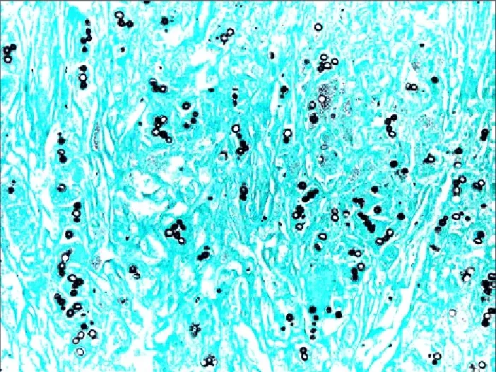
Due to the reduction process of the silver nitrate solution, the fungi species will tint black. After reduction, silver nitrate solution produces black silver ions, which stain fungal cells black.
Due to the conversion of silver nitrate to silver ions, the mycelium and hyphae of the fungus stain a rose pink/pink-red, while the mucin stains a dark grey. Using the light green option will make the background appear quite bluish.
Precautions
- Put on protective gear including lab coats, glasses, and gloves. The fume hood is where you should keep any uncapped hot solutions. You should stay away from any chemicals or dyes that might be around.
- Chromic acid is highly corrosive to the skin and mucous membranes, highly poisonous to the kidneys, and highly carcinogenic.
- Sulfite of sodium Ingestion can lead to stomach pain and irritability, therefore it’s best to avoid it. It’s irritating to the eyes, skin, and other mucous membranes, too.
- The skin and eyes might be irritated by silver nitrate.
- The oxidizer is a tumorigenic substance that, if consumed, can induce severe stomach pain and may be carcinogenic.
- Ingestion of sodium thiosulfate is poisonous and can lead to inflammation of the digestive tract, skin, eyes, and respiratory system.
- In certain cases, exposure to Light Green SF Yellowish has been linked to cancer.
Applications of Grocott-Gomori’s Methenamine Silver Staining
- Fungal infection diagnosis: Grocott-Gomori’s Methenamine Silver Staining is used to diagnose fungal infections, especially those caused by arthroconidia-forming fungi, by specifically visualizing the fungal organisms in tissue specimens.
- Pathology: The staining technique is used in pathological examinations of tissue specimens to identify the presence of fungal infections, which is important for disease diagnosis and treatment.
- Microbial research: The staining technique is also used in microbial research, specifically in the study of fungal infections and their impact on human health and the environment.
- Medical mycology: Grocott-Gomori’s Methenamine Silver Staining is a useful tool in medical mycology, the study of fungi and their impact on human health, as it helps to identify and diagnose fungal infections in patients.
- Environmental monitoring: The staining technique can be used in environmental monitoring to detect the presence of fungal spores in air, water, and soil samples, which is important for understanding the spread of fungal infections and for controlling their impact on human health.
- Quality control in pharmaceuticals: Grocott-Gomori’s Methenamine Silver Staining is used in the quality control of pharmaceutical products to ensure that they are free of fungal contamination, which can have negative impacts on their effectiveness and safety.
- Food safety: The staining technique can also be used in food safety to detect the presence of fungal spores in food products, which can cause spoilage and pose health risks to consumers.
- Agriculture: Grocott-Gomori’s Methenamine Silver Staining is used in agriculture to study the impact of fungal infections on crops and to develop strategies for controlling their spread and impact.
Advantages of Grocott-Gomori’s Methenamine Silver Staining
- Specificity: The staining technique is highly specific and only stains fungal organisms, which makes it an effective tool for the diagnosis of fungal infections.
- High sensitivity: Grocott-Gomori’s Methenamine Silver Staining is capable of detecting even low levels of fungal organisms, which makes it a useful tool for the early detection of fungal infections.
- Easy to perform: The staining technique is relatively simple to perform and does not require complex equipment or special skills, making it accessible to a wide range of laboratories and researchers.
- Cost-effective: Compared to other special staining techniques, Grocott-Gomori’s Methenamine Silver Staining is relatively inexpensive and does not require specialized equipment, making it a cost-effective option for many laboratories.
Limitations of Grocott-Gomori’s Methenamine Silver Staining
- Limited to fungi: The staining technique is only effective for staining fungal organisms and is not useful for visualizing other types of microorganisms.
- False negatives: In some cases, the staining technique may produce false negatives, particularly if the fungal organisms are present in low numbers or if they are masked by other tissue components.
- False positives: In rare cases, the staining technique may produce false positives, in which non-fungal structures are mistakenly identified as fungal organisms.
- Limited sample types: Grocott-Gomori’s Methenamine Silver Staining is typically only used on formalin-fixed, paraffin-embedded tissue specimens and is not appropriate for use on other types of samples.
- Lethal: Skin and stomach irritations can result from inhaling the chemical chemicals. It has been shown that the reagents themselves cause cancer.
FAQ
What is Grocott-Gomori’s Methenamine Silver Staining?
Grocott-Gomori’s Methenamine Silver Staining is a histological staining technique used to identify and visualize fungal organisms in tissue specimens.
What types of samples can be stained with Grocott-Gomori’s Methenamine Silver Staining?
The staining technique is typically used on formalin-fixed, paraffin-embedded tissue specimens.
What are the advantages of using Grocott-Gomori’s Methenamine Silver Staining?
The staining technique is highly specific, sensitive, easy to perform, and cost-effective.
What are the limitations of Grocott-Gomori’s Methenamine Silver Staining?
The staining technique is limited to fungi, may produce false negatives or false positives, and is typically only used on formalin-fixed, paraffin-embedded tissue specimens.
How does Grocott-Gomori’s Methenamine Silver Staining work?
The tissue specimen is first treated with a methenamine silver solution, which is then counterstained with a basic fuchsin stain to enhance the contrast of the fungal organisms.
What is the procedure for performing Grocott-Gomori’s Methenamine Silver Staining?
The procedure typically involves fixing the tissue specimen, embedding it in paraffin, sectioning it, and staining it with methenamine silver and basic fuchsin.
Is Grocott-Gomori’s Methenamine Silver Staining specific to fungi?
Yes, the staining technique is highly specific to fungi and is not effective for visualizing other types of microorganisms.
Can Grocott-Gomori’s Methenamine Silver Staining produce false results?
In rare cases, the staining technique may produce false negatives or false positives.
How does Grocott-Gomori’s Methenamine Silver Staining compare to other staining techniques for fungal diagnosis?
Grocott-Gomori’s Methenamine Silver Staining is a highly specific and sensitive technique that is easy to perform and cost-effective, but it may produce false results in rare cases.
What are the applications of Grocott-Gomori’s Methenamine Silver Staining?
The staining technique is used in fungal infection diagnosis, pathology, medical mycology, microbial research, environmental monitoring, quality control in pharmaceuticals, and food safety.
References
- https://www.reference.com/science-technology/nitric-acid-stain-skin-yellow-9475502370e787b5
- https://www.researchgate.net/post/Which-stains-can-be-used-for-Staining-Fungi
- https://quizlet.com/185455989/histology-stains-flash-cards/
- https://www.clinisciences.com/coloration-de-grocott-colorations-5554/grocott-methenamine-silver-stain-351002431.html
- https://pubmed.ncbi.nlm.nih.gov/14398663/
- https://pubmed.ncbi.nlm.nih.gov/26493433/
- http://www.ihcworld.com/_protocols/special_stains/grocott_methenamine_ellis.htm
